


Analysis Service
PROTIA Inc. provides basic analysis to characteristic analysis services and is committed to satisfying customer needs.
We will do our best to produce the satisfying results through close consultation with customers.

- SERVICES
- Protein Analysis Notice: Undefined offset: 3 in /home/host/proteometech2022/www/html/_skin/layout/inc_navigation_middle.php on line 86
Protein analysis
1. Sample preparation
-
Introduction of the experiment
Service for extracting and separating proteins from sample for protein analysis
Sample precipitation & concentration, Protein quantification, Buffer exchange, removal of redundant protection in blood is provided. -
Precautions for requesting analysis
- In order to extract and separate proteins from the sample, you must remove salt, nuclear acid, polysaccharides, lipid, etc. from the sample. (In particular, salt must be removed using the sample's methanol precipitation, TCA/Acetone precipitation, etc. so that the strip does go wrong during Isoelectric focusing.)
- Special care is required to prevent contamination with Keratin when preparing samples for protein identification using MS. (The contamination of the sample by Keratin has a serious effect on protein identification.)
- After removing the protein band from gel, put it in a 1.5mL tube and send it refrigerated.

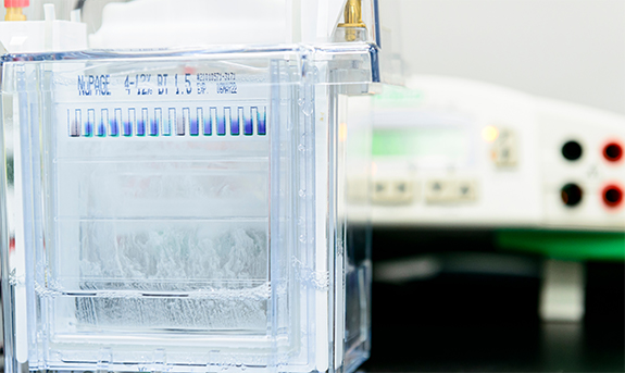
2. One-Dimensional Electrophoresis (1-DE)
-
Introduction of the experiment
Polyacrylamide Gel Electrophoresis service
-
Quantitative comparison between samples, immunoprecipitation (IP), and changes in protein after purification can be confirmed.
3. 1-DE Image analysis
-
Introduction of the experiment
Services provided for quantitative comparison of bands in SDS-PAGE
-
The amount of change is provided.
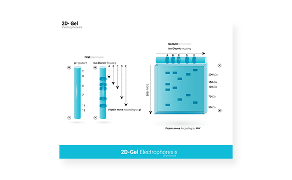
4. Two-dimensional electrophoresis (2-DE)
-
Introduction of the experiment
A service that separates and provides proteins at the desired isoelectric point using an IPG (immunized pH gradient) strip in various pH ranges for isoelectric point separation
-
After primary separation by the isoelectric point, the protein is secondarily separated according to the molecular weight of the protein through Gel% suitable for the sample.
-
It usually lasts 14 ~ 18 hours, and depending on the characteristics of the sample, we provide images by dyeing them in various ways.
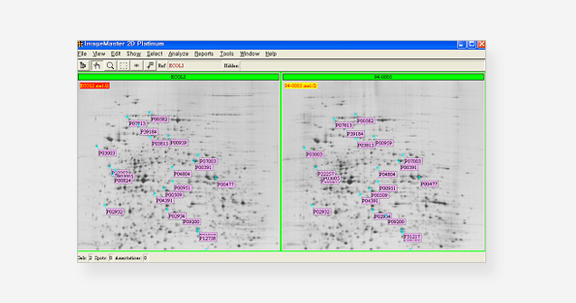
5. 2-DE Image Analysis
-
Introduction of the experiment
By using Image master 5.0 (Amersham Biosciences), a gel image analysis program, provides images and data by analyzing protein spots that show differences in expression in a short period of time
-
Quantitative analysis of changes in all proteins on 2DE, and qualitatively identifying changes to disappearing proteins that cannot be processed by comparison programs.
6. Isoelectric Focusing
-
Introduction of the experiment
Service provided when only the isoelectric value (ispotential point, pI) of a protein is identified
-
It is provided by measuring the pI value of the desired protein bands using IEF Gel (pH 3-10).
7. Gel Staining
-
Proteomtech Inc. makes its own special dye for the experiments. Based on accumulated technical know-how, provides high-resolution images.
-
Coomassie Blue Staining: Produced by Proteomtech, the Comassie Pro (G250) provides a highly-sensitive image.
It's more sensitive than any other product, and you can get a more detailed protein image by giving it a dyeing time of four hours to overnight.
In addition, the destination process is simple during mass spectrometry, saving you valuable time.
Most samples dyed with Coomassie blue can be identified during mass analysis. -
Silver Staining: Modified silver dyeing developed by ProteomTech Inc.
You can check the clean result by removing a lot of background, which is pointed out as a disadvantage of silver dyeing.
It is 10 to 100 times more sensitive than the average comassie blue dye, so it is difficult to identify because a relatively small amount of protein is used for mass analysis.
8. Western blot
-
Introduction of the experiment
Analysis services used to detect specific proteins using antibodies in samples
-
Provided primary antibodies and secondary antibodies is used. If they are not provided, price of primary or secondary antibodies will be charged.
9. Western blot band intensity Image analysis
-
Introduction of the experiment
Service provided for quantitative comparison of detection bands identified after Western blot
-
Provides the intensity of the band as quantitative data.